Modern imaging methods allow us to quickly and accurately diagnose a variety of animal diseases and injuries.


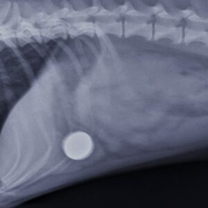

Digital X-ray Examination
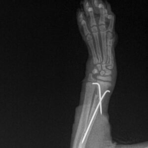
Digital X-ray examination allows us to detect developmental joint defects and quickly diagnose fractures or unusual changes in the chest and abdominal cavity. We have a high-quality AGFA digitizer at our disposal, which displays even subtle changes in bones or soft tissues.Contrast Myelography
Contrast myelography makes it possible to visualize the spinal cord on an X-ray image using a contrast agent injected into the spinal canal. Thanks to this imaging method, it is possible to accurately determine, for example, the location of spinal cord compression and perform a targeted surgical procedure.Ultrasound (Sonography)
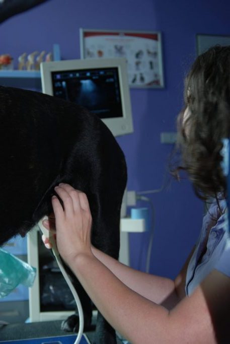
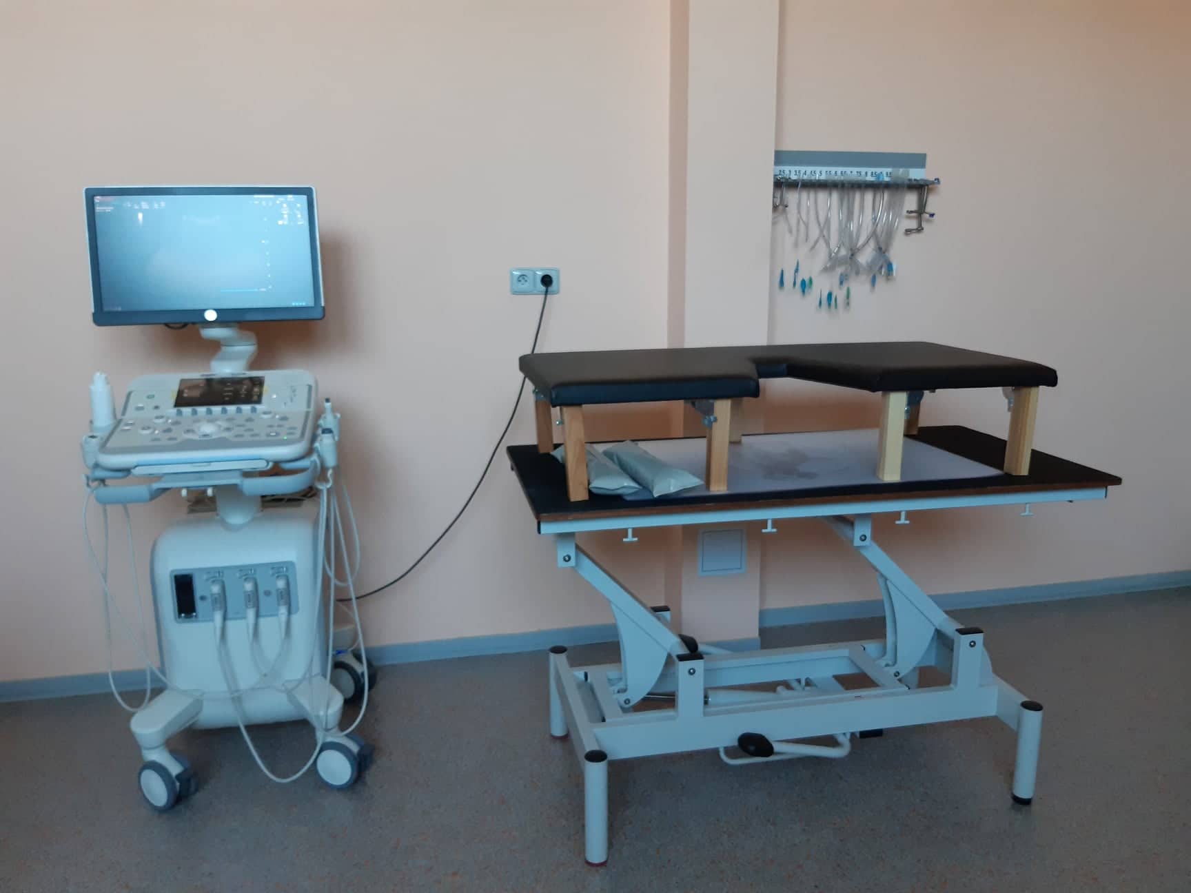
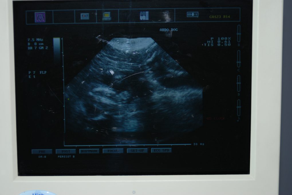
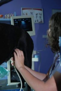
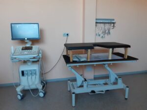
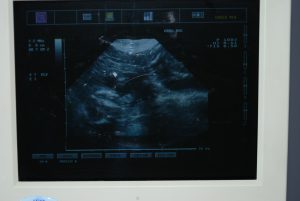
During a sonographic examination of the abdominal cavity, we can detect diseases of the liver, spleen, or intestines. Ultrasound is also suitable for diagnosing heart diseases or confirming pregnancy and monitoring fetal development.Endoscopy
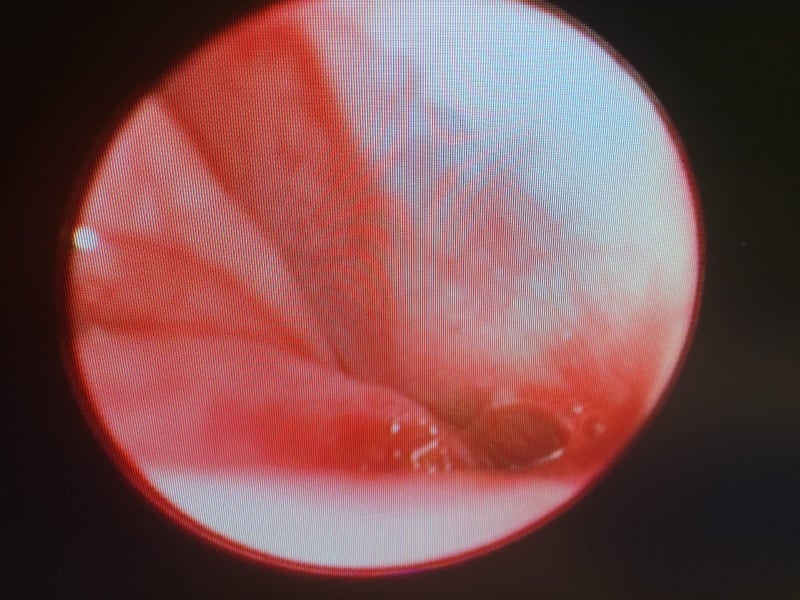
An endoscope allows us to examine the oral cavity, esophagus, nose, larynx, and trachea from the inside, revealing foreign bodies, inflammation, or damaged tissue. The removal of a foreign body takes place using special instruments under camera control.Arthroscopy
Arthroscopy is used to examine joints using special instruments and a camera. It helps us find changes that cause pain to the animal and lead to limping. The removal of damaged cartilage or bone spurs is gentle, without a large surgical wound, and leads to pain relief.